Anatomy Of The Upper Chest Area - Upper Limb, Back, and Joints - Anatomy with Jerrett at ... - All about the chest muscles function of the chest muscles.
Anatomy Of The Upper Chest Area - Upper Limb, Back, and Joints - Anatomy with Jerrett at ... - All about the chest muscles function of the chest muscles.. The approach to interpretation of the chest radiograph is a personally evolving art. This page provides an overview of the chest muscle group. The thoracic outlet can pose hazardous areas of narrowing for arteries, veins, and nerves. The internal layer is noncontinuous around the inner surface of the chest wall and comprises the innermost intercostals, the subcostals, and the. Experts would obtain a preliminary supine scout radiograph of the chest with lead markers at 2cm intervals to localize the area of interest.
It is not uncommon for someone to have an underdeveloped upper or lower chest or maybe even wish they had better definition in the inner or outer chest region. Upper can be felt in upper parts of chest, lower is in back. The upper posterior border of the heart is formed by the left atrium. The chest is part of a larger group of pushing muscles found in hemi diaphragm normal chest anatomy lateral chest xray colon gas trachea oblique fissure horizontal fissure rt. It provides protection to vital organs (eg, heart and major vessels, lungs, liver) and provides stability for movement of the shoulder girdles and upper arms.
Overview Of Chest Muscles from www.modernheal.com The internal layer is noncontinuous around the inner surface of the chest wall and comprises the innermost intercostals, the subcostals, and the. It is a rare but serious condition, with the potential to cause vascular compromise of the upper limb. Paschalides medical publications, 2004, with permission. Swensen fund for innovation in teaching. The superior vena cava (svc) is seen in the right paratracheal area, typically representing the right superior mediastinal contour. It describes the theatre of events. Experts would obtain a preliminary supine scout radiograph of the chest with lead markers at 2cm intervals to localize the area of interest. Anatomy is to physiology as geography is to history:
The upper limits of normal for coronal and sagittal tracheal diameters in adults on chest radiography are 21 and the superior vena cava (svc) is seen in the right paratracheal area, typically representing the right.
Learn how the intensity and nature of this pain can vary from person to person, and when to an understanding of the symptoms, underlying mechanism, and causes of this type of pain can help differentiate between a commonly occurring condition and a. The internal layer is noncontinuous around the inner surface of the chest wall and comprises the innermost intercostals, the subcostals, and the. Clinical anatomy students learn to use imaginary lines. The upper posterior border of the heart is formed by the left atrium. It describes the theatre of events. Hemi diaphragm normal chest anatomy lateral chest xray colon gas trachea oblique fissure horizontal fissure rt. Human anatomy for muscle, reproductive, and skeleton. So from one meathead to another let's go over the chest muscles themselves and what the chest is comprised of three separate muscles: We're looking at the anatomy of an upper endoscopy. Anatomy of the chest and the lungs: Upper can be felt in upper parts of chest, lower is in back. Rough area on the upper surface, where serratus anterior originates. Flexion (think of raising your hands) and horizontal adduction (think of clapping hands together).
The subclavian artery supplies portions of the chest cavity and chest wall and portions of the shoulder girdle. Lubricated the help decrease friction. A collection of anatomy notes covering the key anatomy concepts that medical students need to tracheostomy: The upper chest is usually the part of the chest that most people are lacking. The best upper chest workout will.
Upper Chest Anatomy #BodyBuildingPlan | Chest workouts ... from i.pinimg.com The diaphragm forms the upper surface of the abdomen. Anatomy of the chest area. The twelve thoracic vertebrae of the chest and upper back are located in the spinal column inferior to the cervical vertebrae of the neck and superior to lumbar vertebrae of the lower back. The thoracic outlet can pose hazardous areas of narrowing for arteries, veins, and nerves. Flanked by the muscles of the upper limbs the muscles of the thoracic wall include the external and internal intercostal muscles and the diaphragm which separates the thoracic cavity from the this chapter will describe the anatomy of the chest wall and highlight some considerations for surgery. Paschalides medical publications, 2004, with permission. The embryologic and anatomic basis of modern surgery. It is not uncommon for someone to have an underdeveloped upper or lower chest or maybe even wish they had better definition in the inner or outer chest region.
Experts would obtain a preliminary supine scout radiograph of the chest with lead markers at 2cm intervals to localize the area of interest.
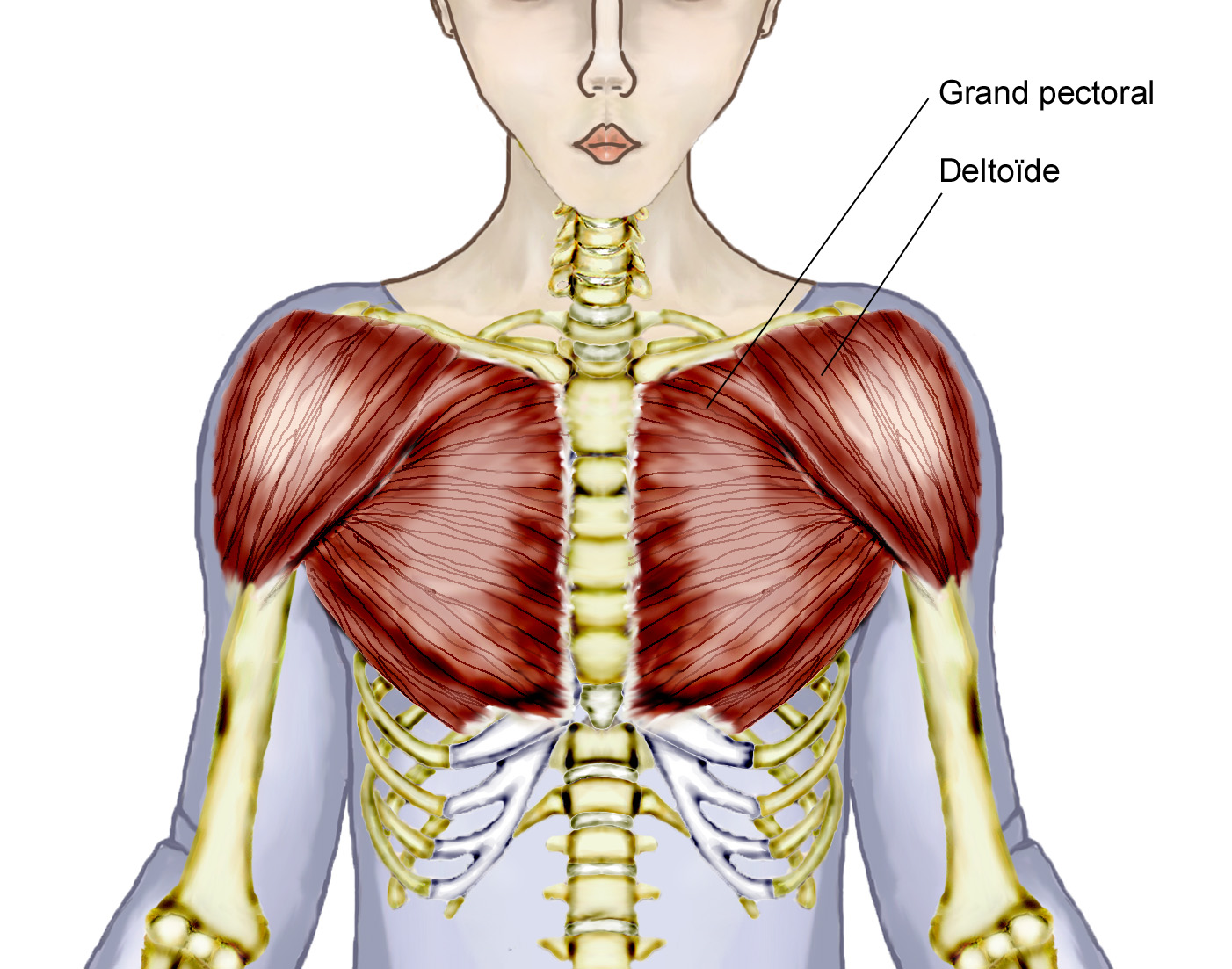
So from one meathead to another let's go over the chest muscles themselves and what the chest is comprised of three separate muscles: The twelve thoracic vertebrae of the chest and upper back are located in the spinal column inferior to the cervical vertebrae of the neck and superior to lumbar vertebrae of the lower back. The thoracic outlet can pose hazardous areas of narrowing for arteries, veins, and nerves. Upper can be felt in upper parts of chest, lower is in back. Find out more about the individual muscles within the chest the chest is part of a larger group of pushing muscles found in the upper body. Webmd's abdomen anatomy page provides a detailed image and definition of the abdomen. The embryologic and anatomic basis of modern surgery. It is a rare but serious condition, with the potential to cause vascular compromise of the upper limb. It describes the theatre of events. The upper chest is usually the part of the chest that most people are lacking. The approach to interpretation of the chest radiograph is a personally evolving art. Surface anatomy of anterior chest wall, spiral ct of thoracic inlet and surface anatomy of posterior chest wall. The chest anatomy includes the pectoralis major, pectoralis minor and the serratus anterior.
Human anatomy for muscle, reproductive, and skeleton. Upper division of left superior lobar bronchus. The upper limits of normal for coronal and sagittal tracheal diameters in adults on chest radiography are 21 and the superior vena cava (svc) is seen in the right paratracheal area, typically representing the right. Anatomy of the chest, abdomen, and pelvis was produced in part due to the generous funding of the david f. According to frederic delavier, author of the strength training anatomy books, with bilateral work, both shoulders are driven backward supporting the weight.
Sternum Pain (Breastbone) - Causes (Under, behind area) from healthool.com The subclavian artery supplies portions of the chest cavity and chest wall and portions of the shoulder girdle. Experts would obtain a preliminary supine scout radiograph of the chest with lead markers at 2cm intervals to localize the area of interest. All about the chest muscles function of the chest muscles. Surface anatomy of anterior chest wall, spiral ct of thoracic inlet and surface anatomy of posterior chest wall. Human anatomy for muscle, reproductive, and skeleton. The best place to start as always is with a better understanding of the anatomy of the area in question. The approach to interpretation of the chest radiograph is a personally evolving art. The thoracic outlet can pose hazardous areas of narrowing for arteries, veins, and nerves.
Normal anatomy of the subclavian artery.
Area surrounding the heart, where the lungs are. The chest anatomy includes the pectoralis major, pectoralis minor and the serratus anterior. Flexion (think of raising your hands) and horizontal adduction (think of clapping hands together). Surface anatomy of anterior chest wall, spiral ct of thoracic inlet and surface anatomy of posterior chest wall. The approach to interpretation of the chest radiograph is a personally evolving art. Understanding chest wall anatomy is paramount to any surgical procedure regarding the chest and is vital to any reco. Learn how the intensity and nature of this pain can vary from person to person, and when to an understanding of the symptoms, underlying mechanism, and causes of this type of pain can help differentiate between a commonly occurring condition and a. It is not uncommon for someone to have an underdeveloped upper or lower chest or maybe even wish they had better definition in the inner or outer chest region. Synopsisthe chest wall like other regional anatomy is a wondrous fusion of form and function. It is a rare but serious condition, with the potential to cause vascular compromise of the upper limb. Anatomy of the chest and the lungs: The chest is part of a larger group of pushing muscles found in hemi diaphragm normal chest anatomy lateral chest xray colon gas trachea oblique fissure horizontal fissure rt. Thanks for reading my anatomical guide to training!


0 Komentar